Monthly Archives: July 2017

Food, Growth and Climate Change

By: Melissa Dellatorre

Hi, my name is Melissa and I am entering my second year as a graduate student in Dr. Donal Manahan’s lab at USC, where I use sea urchins and oysters to study early developmental physiology.

Two major goals of our lab are to increase the efficiency of the aquaculture business by studying influences on growth and development, and to determine how these species are likely to be affected by future global environmental changes such as temperature, food availability, and CO2.

My goals as a WIES Fellow for this summer are to further understand how food quantity and quality affects larval growth at the physiological, biochemical, and whole-organism levels. Although studies have been conducted for decades, there is still a large gap in knowing mechanistically how food is converted into energy, and how the energy budget of an organism is influenced by varying food concentration.

Screen Shot 2017-07-21 at 10.20.23 AM

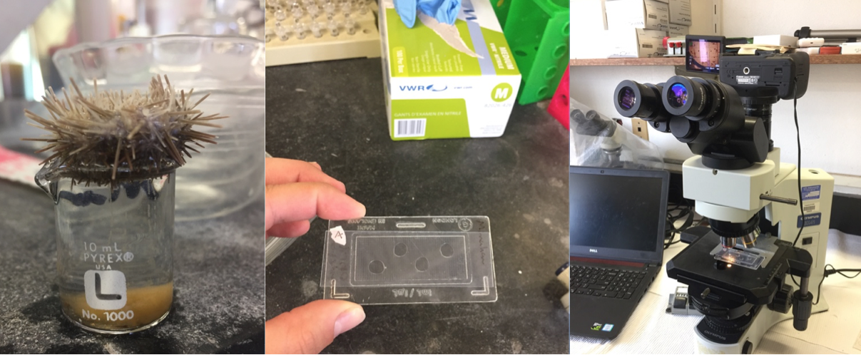
Left: Female Lytechinus pictus urchin spawning ~400,000 eggs into a small beaker. Middle: Aliquots of concentrated eggs for counting to determine total number. Right: Microscope used for counting and taking photographs used to measure size and growth.

This week my labmate Jason and I spawned white sea urchins, Lytechinus pictus, to create two million individuals. This ensures just enough biomass for a two-week experiment, assuming that all of the larvae develop properly. Once spawned, larval sea urchins develop quite rapidly through several phases. By four days old they reach a pluteus stage with developed digestive systems, and are ready to feed.

Screen Shot 2017-07-21 at 10.20.36 AM

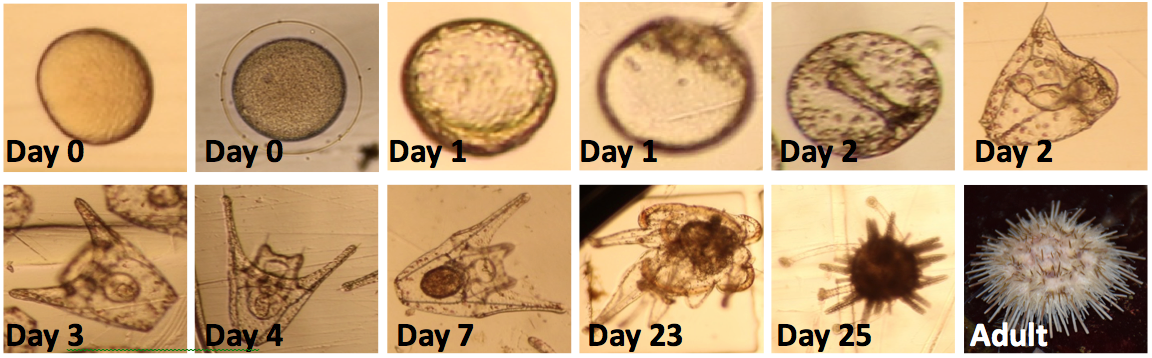
White sea urchin, Lytechinus pictus, development at 15°C. By day 1, organisms enter a blastula stage and begin to gastrulate. On day 2, they move from a gastrula to a prism. On day 3, pluteus stage is reached and their circular stomachs are evident. By day 25 some larvae have reached metamorphosis.

These two million individuals were allocated into six tanks, and are being fed a red alga, Rhodomonas lens, at high, medium, and low concentrations. From here, we take measurements to get the full picture of how these organisms are developing. This includes taking photographs to measure their growth, and doing biochemical analyses of protein, carbohydrate, and lipid contents of the algae and the larvae.

We also measure respiration and protein synthesis rates, as well as clearance rates which tells us how fast they are consuming their food. From this we can determine how many algae have been consumed by the larvae, giving us a measure of intake. This rate also tells us how often we need to top off the tanks with algae to keep food concentrations constant.

Untitled

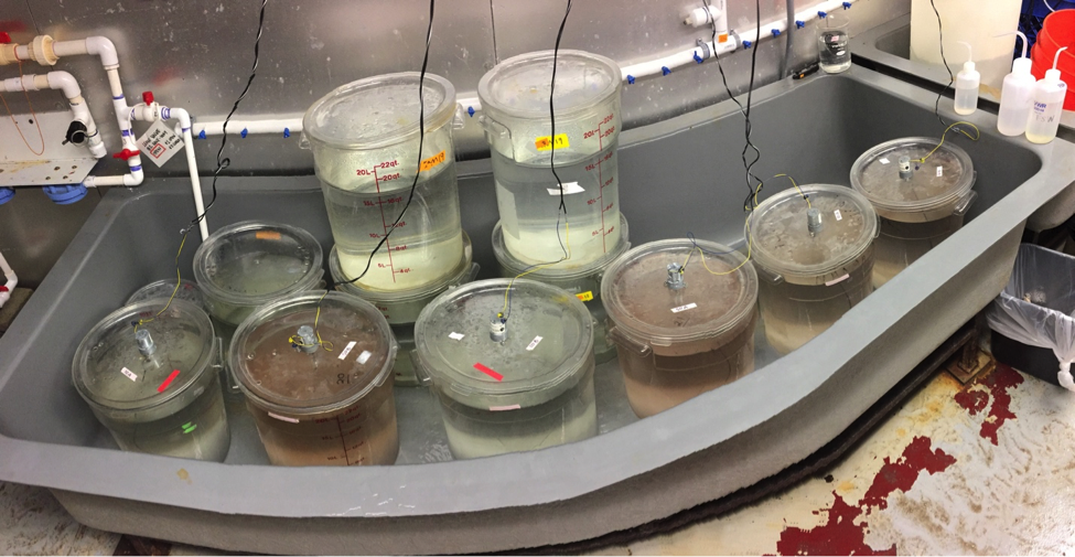
Experimental set up of our six 20 liter tanks (front row). Darker pigmentation can be seen in the tanks with the higher algal feeding concentrations.

My future experiments will compare this alga with other types of algae that white sea urchins like to eat, such as Dunaliella tertiolecta. The results can help shed light on food conversion efficiencies for white sea urchins, and determine the importance of nutrition during larval stages of development.

This research has implications for how larvae will fare under changing ocean conditions that affect food availability. It ultimately may also help to optimize the production of sustainable seafood for society.

Exploring Ocean Microbes

By: Elaina Graham

Hi! I am finishing my second year as a PhD student at USC in Dr. John Heidelberg’s lab. I am a microbial ecologist who looks at genes directly from the environment to explore the diversity of microbial life, and I’m also a WIES Fellow at Catalina this summer.

Microbes truly rule the world (or at least the ocean!) because they help mediate all of the major nutrient cycles in the ocean by helping to turnover carbon, nitrogen, and even sulfur. In previous research, we’ve found a very interesting organism that falls into a group called ‘Aerobic Anoxygenic Phototrophs’ (AAnP) – it uses light to produce energy, but has never before been shown to convert carbon sources like CO2 into organic carbon sources that can be used by the cell. This organism was seen all across the global ocean, in samples we looked at from the North Pacific, North Atlantic, and Mediterranean Sea. Our goal is to determine if it is now present in the waters around Catalina Island.

Because around 99% of the microbes in the ocean haven’t been cultured yet, it is important for us to find other methods to look at the diversity of microbes in the ocean.

Untitled

Aboard the RV Yellowfin, getting ready to collect water. Each bottle on the rosette you see here can collect 12 Liters of water

Spending the summer out at Wrigley has been an amazing experience thus far because it has given me direct access to seawater from the cove that I can use to look for signs of the organism. Because these particular bacteria can make up anywhere from 1% -11% of the community, we often need a lot of water to look for the genes indicating the presence of this group. I have largely been sampling in the cove, but also had a wonderful opportunity earlier in the summer to go on USC’s boat RV Yellowfin (with a class I was teaching out at Wrigley prior to starting my summer research). This helped really kick off my project by enabling me to not just collect surface waters in the cove, but also sample by vessel for water that is 100 meters below the surface!

Once I got this water, I filtered it and began using something called PCR to start looking for signs of AAnP microbes. The method of PCR searches the environmental DNA for a specific gene, and once it finds this gene it amplifies it and produces billions of more copies – enough that we can ultimately sequence. Doing this requires you to be extra clean because even the tiniest bit of contamination from dust in the air or a cough could ruin the experiment. To avoid this we do these experiments in a special clean hood.

Screen Shot 2017-07-17 at 1.48.16 PM

(Left) The PCR clean hood we use to do our molecular work and keep contamination out. (Right) The machine we use to run PCR by cycling through hot cycles up to 96°C.

While doing this I found myself getting very low amounts of ‘positive’ results, which could either have been because A) our group was missing from Catalina waters or B) there were issues with the PCR. Because I had seen previous studies at the San Pedro Ocean Time Series (SPOT) indicating the presence of these organisms, I decided I needed some kind of positive control. So I recreated a classical experiment where multiple AAnP bacteria were isolated from algae – I chose to use Sargassum horneri. AAnP bacteria tend to be pink, orange, or yellow due to the pigments they use to acquire light, so if they grow on solid media they are quickly identified!

Screen Shot 2017-07-17 at 1.49.39 PM

(Left) My incubator setup for culturing bacteria. I have solid agar plates where I isolated pigmented bacteria from the algae, as well as liquid media where I am using algae to grow up a culture of its associated bacteria. (Right) Close up of some of these cultures

In addition to my research I have been getting to do a lot of education and outreach with some awesome groups! The most recent was the USC C-DEBI high school group who wanted to isolate some bioluminescent bacteria from the cove (see picture below). They were so excited to learn about marine microbes and were very cool to work with! I took a couple of days to go back to the mainland after they left, and now I am looking forward to starting new experiments this upcoming week to continue my search for this novel AAnP group!

Screen Shot 2017-07-17 at 1.49.54 PM

(Left) A picture of bacteria I helped some high school students put on a plate to isolate bioluminescence. (Right) A picture of the same plate with the lights out showing the bioluminescenct colonies.